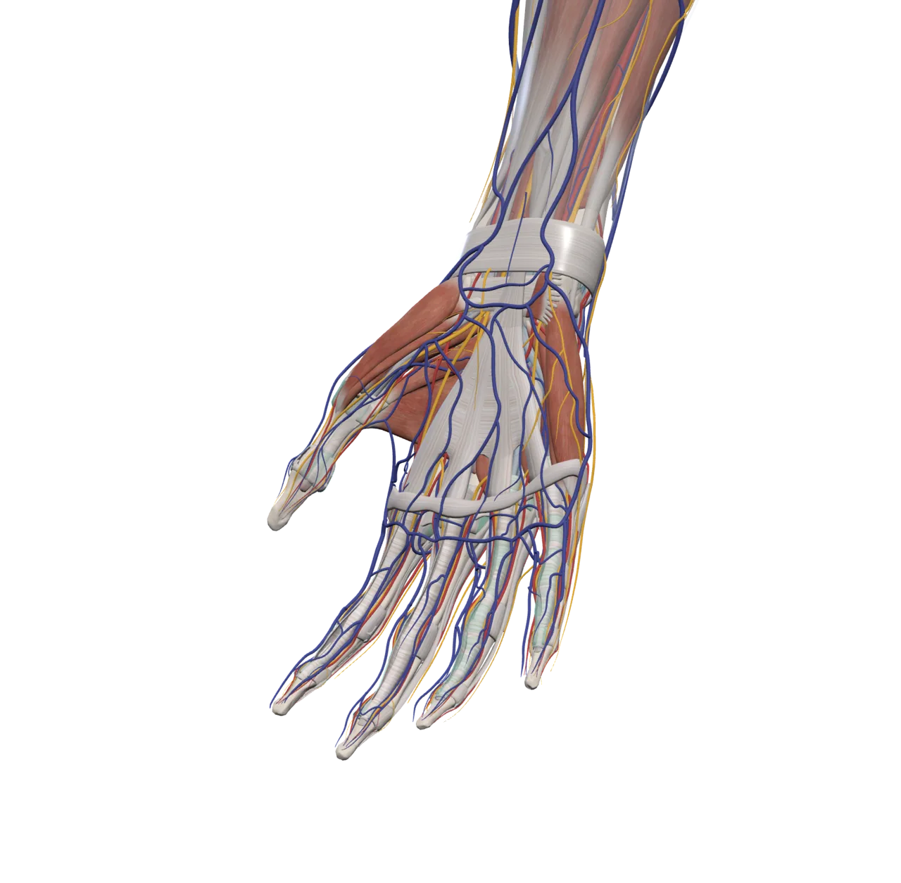

Explore the full body in 3D
Navigate every system, region, and structure in context, from the whole body down to a single nerve.
Ask and understand
Get clear answers about what you're looking at, how structures relate, and why it matters clinically.
Study at your own pace
Search, isolate, and revisit anatomy as many times as you need. No cadaver lab required.

Practice before the OR
Work through surgical access steps like incision, exposure, and retraction on realistic 3D anatomy.
Build procedural confidence
Repeat workflows until the sequence feels natural, without risk to a patient.
See what you're getting into
Understand the layers, landmarks, and relationships you'll encounter before you pick up a scalpel.

Bring anatomy to life
Pose the body, animate movement, and show how structures behave, not just how they look.
Overlay on the real world
Use AR and live camera tracking to place anatomy on a real person or in a physical space.
Create visuals that teach
Build medical illustrations, explainers, and presentations directly from the atlas.

Open studies locally
Load DICOM files or folders from your device while keeping imaging data in the browser session.
Inspect series and slices
Move through CT or MRI studies with the essentials needed to understand the imaging stack.
Connect images to anatomy
Use DICOM review as the foundation for image-to-atlas workflows and clinical visualization.
Explore the atlas
Built for medical students, clinicians, and developers who need fast, intuitive and visual access to human anatomy.